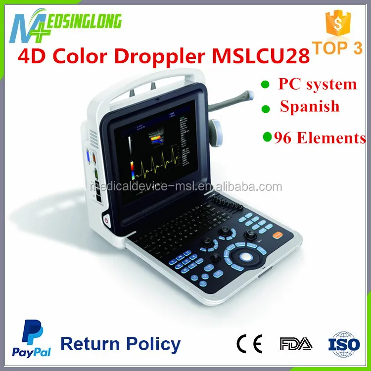
File with a.4DV filename extension contains ultrasound data, generated with the GE Healthcare Voluson dedicated device. It can be opened with the specialised GE View 4D software or ViewPoint, provided the 4D View add-on is installed. Image viewing software can be used by physicians to optimize, analyse and process data obtained via ultrasound.

A 3D ultrasound of a human fetus aged 20 weeks3D ultrasound is a technique, often used in fetal, cardiac, trans-rectal and intra-vascular applications. 3D ultrasound refers specifically to the volume rendering of ultrasound data and is also referred to as 4D (3-spatial dimensions plus 1-time dimension) when it involves a series of 3D volumes collected over time.When generating a 3D volume the ultrasound data can be collected in four common ways.
Freehand, which involves tilting the probe and capturing a series of ultrasound images and recording the transducer orientation for each slice. Mechanically, where the internal linear probe tilt is handled by a motor inside the probe. Using an endoprobe, which generates the volume by inserting a probe and then removing the transducer in a controlled manner. The fourth technology is the matrix array transducer that uses beamsteering to sample points throughout a pyramid shaped volume. Contents.Risks The also apply to 3D Ultrasound.
Essentially ultrasound is considered safe. While other imaging modalities use radioactive dye or ionizing radiation, for example, ultrasound transducers send pulses of high frequency sound into the body and then listen for the echo.In summary, the primary risks associated with ultrasound would be the potential heating of tissue.
The mechanisms by which tissue heating and cavitation are measured are through the standards called thermal index (TI) and mechanical index (MI). Even though the FDA outlines very safe values for maximum TI and MI it is still recommended to avoid unnecessary ultrasound imaging. Applications Obstetrics 3D US is useful among other things for facilitating the characterization of some congenital defects, such as skeletal anomalies and heart issues. With real-time 3D US the fetal heart-rate can be examined in real-time Cardiology The application of three-dimensional ultrasound in cardiac treatments has done outstanding progress in scanning and treating the heart issues. When the 3D ultrasound technology is used to visualize the cardiac state of an individual, it is called as 3D Echocardiography. With the integration of other technologies with the Ultrasound, it is now possible to track the quantitative measures like the chamber volume that occurs during the cardiac cycle.
Also, it provides other useful information like tracking the blood flow, speed of contractions and expansions. With the 3D Echocardiography method doctors now can easily detect the artery diseases and can finely examine the various defects. The echo applications helps to give a real-time image of the cardiac structures. Surgical guidance Traditionally, with the 2D ultrasound, the specific position of organs and tissues useful in surgeries could not be located especially in the oblique plane.
However, with the advent of 3D ultrasound, the imaging technique has evolved manifolds enabling the surgeons to get a real-time picture of the tissues and organs and visualize the complete scan more efficiently. In addition to this, 3D ultrasound provides surgical guidance in terms of treating transplant and cancer with its technique of rotational visualizing while scanning. This technology has evolved various methods like rotational scanning, slice projection, use of integrated array transducer that helps the surgeons to handle the traumatic cancer patients. Further, with 3D ultrasound doctors can now treat various kind of tumors as every tissue can be easily diagnosed and introspected to study the defects and cause. Thus, we see that the ultimate scanning and visualization done by 3D ultrasound has developed better ways of treating the patients with problems like cancer, tumors and transplants.Vascular imaging Blood vessels and arteries movements are not easy to track because of their distribution due to which capturing a real-time picture becomes difficult. Diagnosis is widely used in every kind of treatment and with the 3D US, it is now possible to track dynamic movement of blood cells, veins and arteries.
In addition to this, various types of diagnosis like measuring the diameter, diagnosing the wall between the arteries can be detected with a magnetic tracker integrated with 3D US that helps in accurate positioning. So the technology gives imaging assistance and also has a sensor that helps in tracking the position of vessels in operations.Regional anesthesia Real-time three-dimensional ultrasound is used during peripheral nerve blockade procedures to identify relevant anatomy and monitor the spread of local anesthetic around the nerve. Peripheral nerve blockades prevent the transmission of pain signals from the site of injury to the brain without deep sedation, which makes them particularly useful for outpatient orthopedic procedures. Real-time 3D ultrasound allows muscles, nerves and vessels to be clearly identified while a needle or catheter is advanced under the skin. 3D ultrasound is able to view the needle regardless of the plane of the image, which is a substantial improvement over 2D ultrasound.
Additionally, the image can be rotated or cropped in real time to reveal anatomical structures within a volume of tissue. Physicians at the Mayo Clinic in Jacksonville have been developing techniques using real time 3D ultrasound to guide peripheral nerve blocks for shoulder, knee, and ankle surgery. References.
2 extension(s) and 0 alias(es) in our database
Below, you can find answers to the following questions:
- What is the .4dv file?
- Which program can create the .4dv file?
- Where can you find a description of the .4dv format?
- What can convert .4dv files to a different format?
- Which MIME-type is associated with the .4dv extension?
4D View Ultrasound Data
4DIM Interactive Animation

Other types of files may also use the .4dv file extension. If you have helpful information about .4dv extension, write to us!
Is it possible that the filename extension is misspelled?
We found the following similar extensions in our database:
The .4dv filename extension is often given incorrectly!
Paint tool sai pencil brush downloaded download. According to the searches on our site, these misspellings were the most common in the past year:
Can't open a .4dv file?
If you want to open a .4dv file on your computer, you just need to have the appropriate program installed. If the .4dv association isn't set correctly, you may receive the following error message:
Windows can't open this file:
File: example.4dv
To open this file, Windows needs to know what program you want to use to open it. Windows can go online to look it up automatically, or you can manually select from a list of programs that are installed on your computer.
Welcome to HPE's interactive Citrix XenServer Support and Certification webpage for ProLiant and BladeSystem Servers (x86).Please Note:Citrix XenServer and Hypervisor are Supported and Certified with in-distro drivers only. Hp proliant bl460c g7 driver for mac download.
To change file associations:
- Right-click a file with the extension whose association you want to change, and then click Open With.
- In the Open With dialog box, click the program whith which you want the file to open, or click Browse to locate the program that you want.
- Select the Always use the selected program to open this kind of file check box.
Supported operating systems
Windows Server 2003/2008/2012/2016, Windows 7, Windows 8, Windows 10, Linux, FreeBSD, NetBSD, OpenBSD, Mac OS X, iOS, Android